Diagnosing Back Pain: Where the Challenges Lie
Our new colleague, Robert Savala, MD has been a pain management specialist practicing in the Bay Area for 25 years. In a series of two posts, he will draw on his decades of experience in spinal diagnostics and spinal injections to explain the challenges of accurately diagnosing lower back pain and the latest advanced treatments.
“Look well to the spine for the cause of disease.” -Hippocrates
Even 2,500 years ago, the “Father of Medicine” understood the importance of the spine in our overall wellbeing.
In many ways, he was right. We know today that lower back pain (LBP) is the leading cause of disability in working-age adults in the USA and worldwide. One in five of us will have at least one episode of prolonged LBP, and nearly one in ten will suffer from chronic LBP.
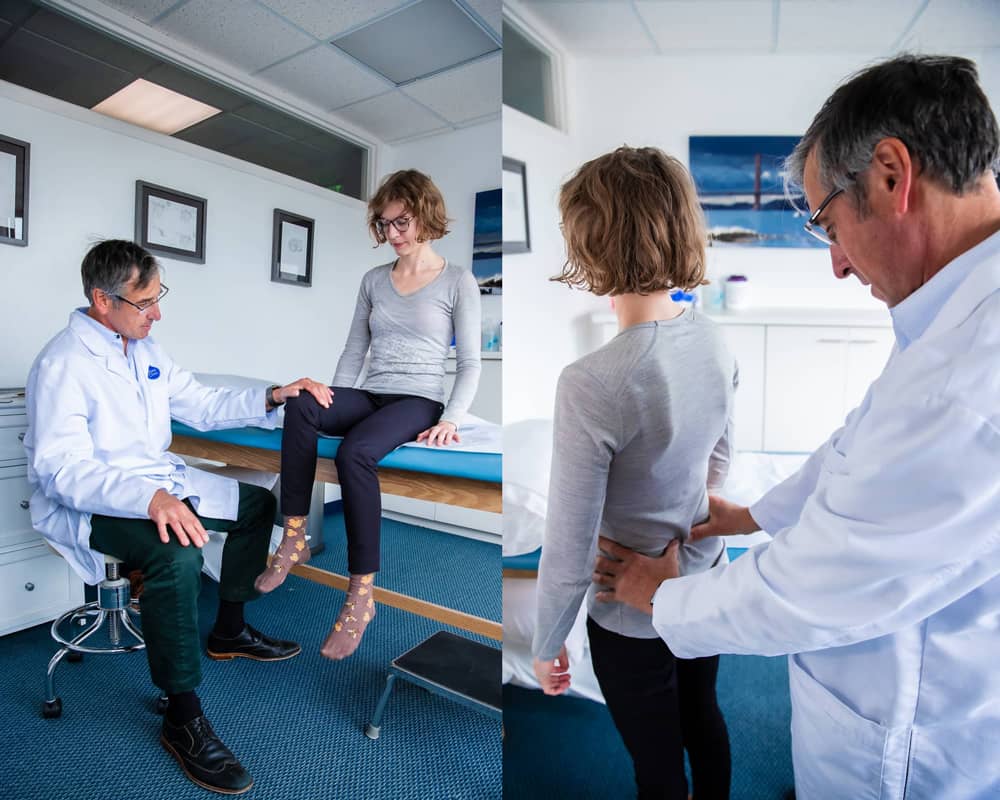
One of the most frustrating aspects of lower back pain is our failure to properly diagnose its source. I hear stories every day from patients who have seen multiple medical professionals who diagnose differing sources of pain and can’t agree on the best course of treatment. One major issue leading to confusion is that spine degeneration often causes pain that radiates to areas beyond the spine. Like an echo in a canyon, it may be generated in one place and experienced in another. Imaging studies are helpful in diagnosis, but far from conclusive. We can’t see pain on an X-ray or MRI. Not everything that looks bad on an image is painful, while areas that appear normal may be sources of pain.
Despite these challenges, we do have a handle on how to approach the spine. An exam provides initial clues as we look for neurological changes that can help us trace the actual source of pain. The patient’s history and imaging also help us make the diagnosis. Targeted spinal injections—my area of expertise—can also help locate the pain source: Relief of pain after an anesthetic injection to a specific structure of the spine can isolate the symptomatic culprit and guide treatment.
Briefly, three structures—all related to the moving parts of the spine—cause the majority of LBP: the facet joints, intervertebral discs (IVD), and sacroiliac joints (SI).
The intervertebral disc (IVD) is a very special tissue. A fibrous ring surrounds a more gelatinous core that absorbs and dissipates pressure. It is designed to act as a shock absorber. Through injury or prolonged static loading (e.g. bending, prolonged standing, or sitting), the IVD begins to degenerate. On a cellular level, the nucleus of the disc loses its ability to hold onto the water that creates the “cushion” of the disc. On the MRI we literally see it shrink, dry out, and bulge.
These changes lead to the disc becoming less able to handle mechanical loads. As the disc begins to fail, there a breakdown of the connective tissues in the disc, creating an intense inflammatory reaction. Biochemical studies show that a damaged disc produces a great number of inflammatory proteins in our body, turning pressure into pain and making our spine less tolerant to loading and static pressures.
Other tissues in our bodies—like skin and bone—regenerate. But the IVD is the largest tissue without a direct blood supply. There are no arteries or veins in discs, limiting the body’s ability to deliver healing nutrients and repair cells. So even minor degeneration can cause a disproportionate amount of pain.
Another source of LBP is facet joints. These connect the vertebrae bilaterally, allowing for additional spinal support and mobility. These, like all joints, can wear down, leading to arthritis and joint overgrowth. As it’s very hard to distinguish between a disc or facet pain source, targeted injections help us diagnose which structures are causing pain. Fortunately, we have simple nerve ablation techniques that can provide relief by destroying the sensation from these joints.
The sacroiliac joint is a third possible culprit. SI joint issues commonly present with pain off-midline in the sacrum and often radiate to the hip—sometimes even to the pelvis and proximal thigh. They are diagnosed by a patient experiencing tenderness over the joint when maneuvers that stress the joint. Imaging can identify joint changes, but a painful SI joint often shows no particular degeneration. So, the gold standard of diagnosis is targeted sacroiliac joint injections and a patient’s response to them.
As LBP is such a drain on so many people, accurate diagnosis is of course essential. But once a diagnosis is made, what are the best possible treatments? I’ll talk about that in my next post.
