Failed Achilles Tendon
Hear From Our Patients
Double Achilles Injury Patient Compares Percutaneous Repair to Open Surgery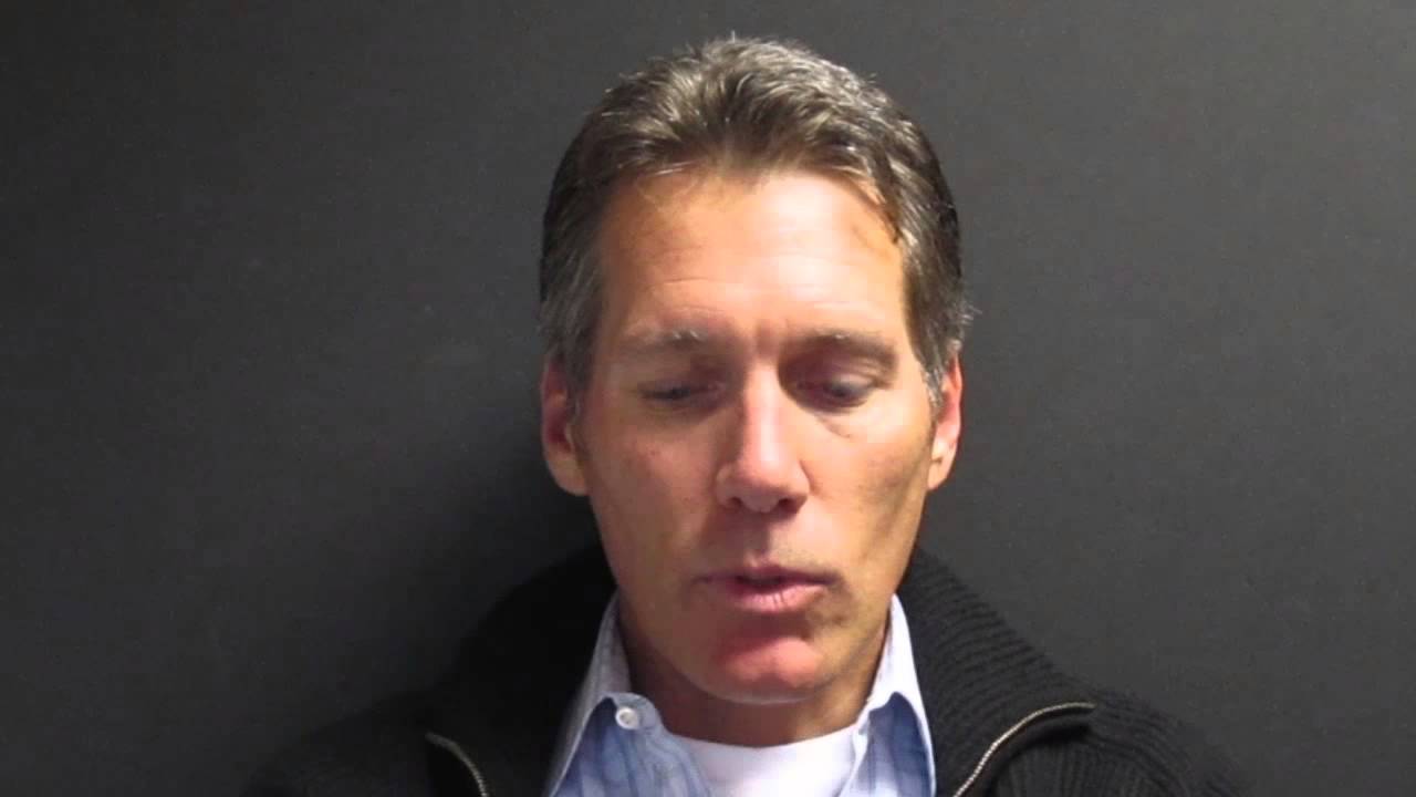
The Achilles tendon stretches from the calf muscles to the heel. It ruptures most commonly in 40-60-year-old male weekend warriors, often with the sound of a pop or a crack. Most of the time, surgical repair works well. Sometimes, though—too often—it goes awry. Here is why.

The tendon is a broad band of strong collagen fibers, surrounded by a thin sheath. The sheath permits the tendon to glide and stretch smoothly. In normal life, we take 2-3 million steps per year, and in order to push our feet off the ground those tendons must elongate as the calf muscles contract. This repetitive cycling permits not just walking, but running and jumping as well—with forces that can exceed five times the person’s body weight.
For some reason, males (and it is almost always males, with a few female ruptures thrown in each year for variety) report that, when they reached for a tennis ball or landed from a basketball shot, they heard what they thought was an actual shot: the bursting of the tendon fibers. Collapsing to the ground with pain, they almost always know, clearly or intuitively, what has happened.
When presenting to their orthopaedic surgeon, a diagnosis is easily made by palpating the gap in the tendons. An MRI helps determine if the tendon itself is diseased or if it just was the unlucky rupture of a middle-aged weekend warrior. A few things can increase the likelihood of rupture, including the use of fluoroquinolone antibiotics (Cipro being the most common), recent chronic tendinitis (made worse by the ill-advised injection of a cortisone shot into the tendon), or chronic high-dose anabolic steroids.
The surgeon who prefers to open these tendons and repair them with sutures takes on a host of new risks. First, the skin and sheath covering the tendon must be incised. Often, the sheath is too thin to repair. The blood clot that naturally forms around the torn ends of the tissues is washed away in the open surgery. This means that all the natural growth factors and stem-derived self-repair cells—which have migrated to the torn ends to initiate a natural repair—are lost. The repair, skin closure, and rehab program all help determine how well the tendon will heal. Unfortunately, with open surgery, there is an increased risk of infection and skin healing problems. These sometimes leave the patient with an angry scar and weakened tendon. Any immobilization has been shown to encourage the formation of scar tissue, rather than normal collagen tendon healing. For this reason, a rehab program must be started on day one, with early mobilization of the repaired tissues. Casts have been abandoned, due to the inability of moving the healing tendon when it is confined.
To minimize these risks, a percutaneous repair technique was developed by Griffen and Ma in 1977, and modified by us and others over time. Without opening the skin, resorbable sutures can be passed through tiny punctures and woven back and forth, pulling the tendon ends together without losing the natural blood clot or compromising the sheath. The results are the same as a successful open repair but without the additional risks. Both techniques can compromise a sensory nerve that runs right over the tendon at the most common site of rupture. But this usually resolves on its own—especially when resorbable sutures have been used.
Failures of percutaneous repairs are exceedingly rare unless a new injury (such as a fall down the stairs) occurs during the post-op period. This is because the Achilles tendon will actually heal on its own if left unrepaired; the additional sutures only optimize the healing environment. Without intervention, a tendon heals with scar tissue bridging the gap between the two tendon ends. This results in a healed but lengthened tendon, which loses some of its strength and push-off power. Percutaneous sutures bring the tendon ends together, permitting healing at the normal length. Almost all athletes choose repair.
The complications of failed repairs are often due to weakness, scar formation, infection, or re-rupture. Salvaging the failures usually does require an open surgery, with a donor graft to rebuild the damaged or missing tissue. It is clearly far better to do the repair right the first time and optimize the patients’ rehab program with the goal of returning to sports fitter, faster, and stronger than they were before “the shot heard round the court.”
To find out more about how you may have your Achilles tendon repaired without open surgery you may download our "Saving My Ankle Guide" here.
