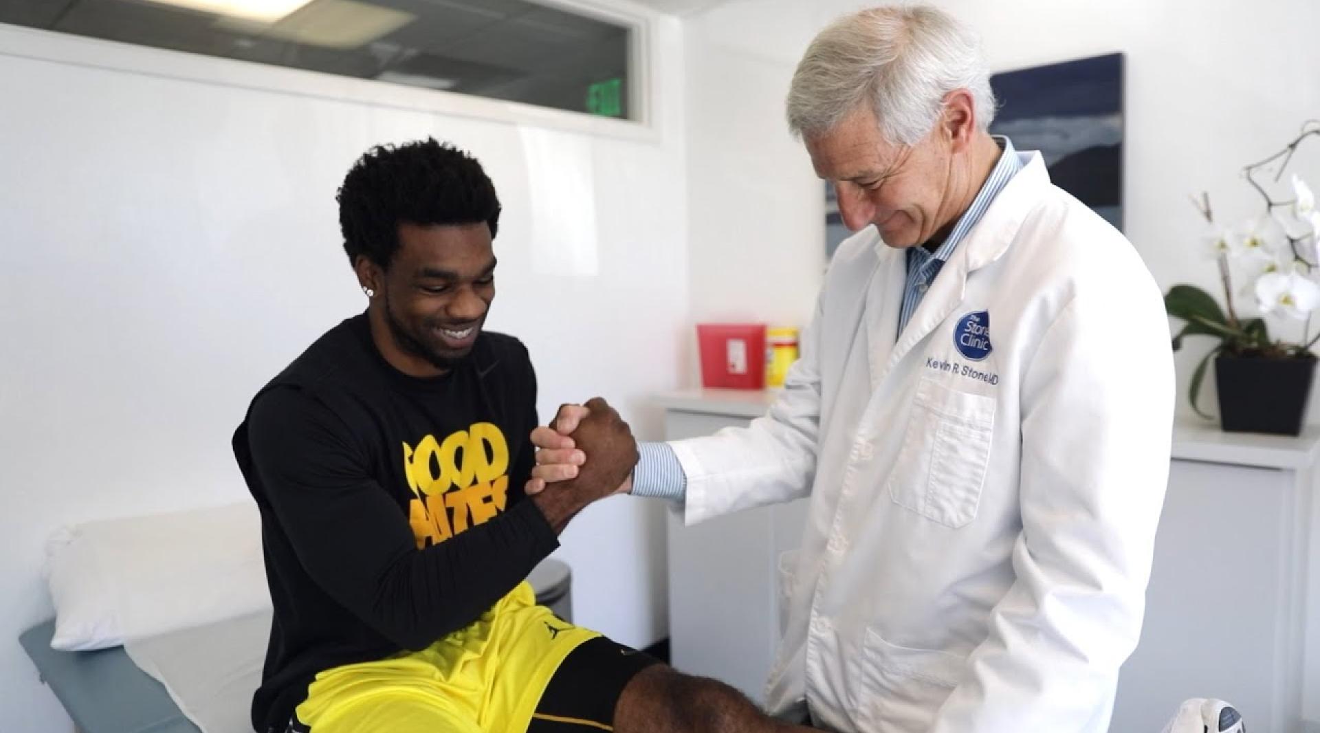
Injury
to the cartilage of the knee joint is one of the most common orthopaedic presentations.
Few orthopaedic problems have received more attention in recent years than the
latest advances in cartilage repair, regeneration and replacement. Surgical procedures
have advanced to such a stage that patients now expect there to be alternatives
to artificial joint replacement in the treatment of their arthritic condition.
Patients also recognize the techniques of yesterday will be improved upon tomorrow,
and question whether to delay current treatment options in anticipation of promising
new ones. A
number of the newer surgical techniques being utilized have shown promising results
in short-term treatment of cartilage dysfunction in the knee. This, as well as
an improved understanding of post-operative treatment programs has restored function
and diminished symptoms in an increasingly large number of people. However, for
many of these newer techniques, there is a lack of long-term follow-up.
This chapter
presents a brief description of structure, biomechanics, evaluation of articular
cartilage injuries and presents some of the surgical techniques designed to stimulate
cartilage repair and regeneration. ARTICULAR
CARTILAGE Articular
cartilage covers the end of bones in joints. The smoothness and thickness of the
cartilage determines the load-bearing characteristics and mobility of joints.
Lesions on the chondral surfaces of joints interfere with the smooth motion of
the joint. This type of damage can cause mild to significant symptoms of pain,
instability and stiffness. Two and a half centuries ago, Hunter first recognized
that damaged articular cartilage does not have the capacity to repair itself.1
This observation has led to a wide variety of treatment approaches for focal chondral
defects in the knee with varying levels of success. Treatments such as drilling,
abrasion, microfracture, and debridement have been shown to provide symptomatic
pain relief and improved function,2-24
although there are very few prospective controlled comparative studies. Most of
these techniques have shown the repair process is dominated by the production
of fibrocartilage instead of normal hyaline cartilage. The fibrous component of
the repair is believed to be produced by fibrocytes carried to the repair site
by blood introduced to the lesion by surgical or traumatic means. Other treatment
options such as periosteal grafting, osteochondral autografts and allografts,
and autogenous chondrocyte cell transplantation2-4,
6, 13,
14, 22,
25-35 have
also shown promising results in reduction of pain and dysfunction and will be
discussed further. Normal hyaline articular cartilage has never been successfully,
reliably reproduced by any technique to date. ANATOMY
Articular
cartilage is a thin, smooth, low friction gliding surface with a remarkable resiliency
to compressive forces. It is a material only a few millimeters thick yet with
excellent wear characteristics. Its mechanical and structural capacity is dependent
on the integrity of its extracellular matrix. Chondrocytes sparsely distributed
throughout a matrix of structural macromolecules work together with a hydrated
extracellular glycosaminoglycans to attract and then sequentially extrude water.
Extracellular components of collagen, proteoglycans, noncollagenous proteins and
water to provide the shear, compressive, and permeability characteristics of cartilage.9,
14, 36-39
This charged mechanical interaction permits cartilage to perform its mechanical
functions without appreciable wear.9,
36-38 It
is the composition and highly complicated interaction of these components that
makes regeneration and replacement techniques challenging. Water
constitutes between 65-80% of the entire wet weight of articular cartilage36,
37 and is
about 15% more concentrated at the surface than in the deeper zones.36
Chondrocyte cells produce the extracellular matrix. Distributed throughout
the matrix, chondrocytes compose less than 5% wet weight and are the derivative
of pleripotential mesenchymal cells that are able to give rise to bone, fat, skin,
cartilage, and tendon.31,
36-38
Collagen
makes up about 15-22% of the wet weight and contains 90-95% type II collagen fibers
with a small percentage of types IX and XI.36-38
This is what provides the high tensile stiffness, strength and resiliency of the
tissue. Proteoglycans constitute about 4-10% of the total wet weight and
are a mix of large aggregating (50-85%) and large nonaggregating (10-40%) proteoglycans.36,
38 They
are responsible for pressure elasticity and charged interactions with water.9,
40 Noncollagenous
proteins, elastins, integrins and other macromolecules of protein are responsible
for the matrix organization and maintenance.37
The functions of articular cartilage include load transmission and distribution,
smooth articulation, and aid in lubrication.14,
30, 36
Load transmission and distribution is due to the ability of the structural matrix
to deform, which leads to increased joint contact areas and distributed mechanical
stresses.36
It also has the ability to respond to applied loads through fluid exudation and
redistribution within the interstitial tissue. HEALING
AND VASCULARITY The
combination of the lack of blood supply and a few cells distributed widely amongst
a dense extracellular matrix leads to a limited ability to heal.1,
6, 14,
15, 36
The usual inflammatory response of hemorrhage, formation of fibrin clot, cellular
production and migration of mesenchymal cells is absent.14,
36 Other
factors such as age, depth and degree of damage, traumatic or chronic condition,
associated instability, previous total meniscectomy, malalignment, and genetic
predisposition are also factors affecting healing of cartilage.6,
31, 36,
37 Age affects
healing in part because in newborns, the multi-functioning mesenchymal stem cells
needed for healing account for 1 in every 10,000 cells in bone marrow and reduces
to 1 in 100,000 in teens, 1 in 400,000 by age 50 and 1 in 2 million in an 80 year
old.31
Depth of the lesion is a factor in healing because surface defects that do not
penetrate the subchondral bone have to rely on sparsely populated chondrocytes
for matrix remodeling where deeper lesions may introduce a blood supply from the
well-vascularized subchondral bone. With the blood comes fibrocytes that modulate
to fibrochondrocytes. These cells produce a relativley disorganized lattice of
collagen fibers partially filling the defect with structurally weak tissue. Traumatic
isolated lesions typically heal better than areas with more degenerative, global
defects. Structural instability and/or other associated pathology also cause uneven
and often excessive forces onto the articulating surfaces.38,
41-43
EVALUATION
A
careful assessment and clinical evaluation of the injured area is critical for
accurately diagnosing an injury to the articular surfaces. Some of the more important
components to consider when evaluating a cartilage injury are: Was it of acute
and/or traumatic nature? Are there other predisposing factors? Is it a chondral
or subchondral lesion? Is there any associated osteoarthrosis indicating progressive
loss, attempted repair, and structural remodeling? Are there indications of degenerative
osteoarthrosis or indications of chronic changes from overuse and/or malalignment?
Was there other pathology created at the time of injury? Terry
was the first to describe the incidence of isolated chondral fractures in 1988.16,
44 With
the advent of the MRI and improved techniques, chondral or subchondral bone bruises
are seen in about 80% of acute anterior cruciate ligament ruptures.14,
45 Whether
or not these bone bruises represent forces that have exceeded the lethal threshold
for the overlying chondrocytes and therefore will lead to eventual frank lesions
is unknown. Patients typically complain of nonspecific episodes of catching, locking,
joint-line pain and low-grade swelling.14,
46 Pain
is not the primary subjective complaint unless the lesion has penetrated subchondral
bone or there is associated synovial irritation.4,
6, 14
Symptomatic catching on the chondral edges, degradative debris and distension
of the joint causing synovitis contributes to pain production.4
Obtaining a careful history, noting the exact mechanism and other predisposing
factors is essential. However, though some studies note a 94% correlation of specific
incidence and tenderness to palpation,16
others have found only a 25-40% correlation of episode and joint line tenderness.47
X-rays
and MRI can aid in improving the diagnostic findings. Sensitivity and specificity
of MRI techniques have improved in detection of chondral lesions greater than
3 mm in diameter.45
MRI has a low sensitivity for chondral delamination injuries of about 21%.47
MRI is very operator and field strength sensitive and is a function of the pulse
sequences and imaging technique used.14
X-rays are preferred for documenting sclerotic changes, osteophytic formation,
compromised joint space (in total and non weight bearing positions), and angle
of alignment. CLASSIFICATION
Classifying
chondral lesions is difficult, frustrating and probably totally unreliable. The
most common classification system used is the Outerbridge system,17
developed as a means for assessing chondral damage to the articular surface of
the patella. There are five levels of degeneration: Grade 0=normal articular cartilage;
Grade I=softening and swelling; Grade II=partial thickness and early fissuring
on the surface, <½ in. in diameter; Grade III=fissuring to the level
of subchondral bone, but the bone not visibly exposed, >½ in. in diameter;
Grade IV=erosion down to subchondral bone.17
However, there is very poor interobserver correlation between surgeons in describing
cartilage lesions. Also, many lesions are a mixture of types. Surface only classifications
fail to consider the underlying damage seen by MRI. TREATMENT
Treatment
of articular cartilage defects in the knee has been attempted in numerous studies
and all with varying levels of success.2-12,
13-16, 19-24
Success of these approaches has generally diminished over time possibly due to
the formation of fibrocartilage, inadequate development of repair tissue, poor
cell differentiation and poor bonding to the surrounding articular cartilage borders.4,
12, 48
Although these techniques can result in symptomatic pain relief and improvement
in functional status, the long-term results remain mixed. When comparing different
conventional therapeutic options, it is important to recognize the goal of surgical
intervention. Treatment can be directed at either treating the symptoms or trying
to affect articular repair or regeneration.4
Repair refers to the restoration of a damaged chondral surface with new tissue
that resembles but does not duplicate the structure, biochemical makeup, function
and durability of articular cartilage. Regeneration denotes the formation of new
tissue indistinguishable from normal articular cartilage.6,
35, 36,
49 Several
investigators have also reported the efficacy of continuous passive motion (CPM)
use following these techniques in improving the visual and chemical appearance
of the defect.20,
21, 36,
43
There is
also the component of other associated pathology in conjunction with focal articular
cartilage damage. Personal observations and select studies have suggested that
isolated chondral defects heal and recover slower functionally than those combined
with other ligamentous injury or surgery.50
This could probably be attributed to the increased cellular activity and cytokine
stimulation with combined lesions.50
A few of the more common treatment methods are noted below. Lavage--Lavage
rids the knee of loose articular debris and inflammatory mediators that are known
to be formed by damaged synovial joints. Jackson had found a 45% symptomatic improvement
in patients at 3½ years. When arthroscopic lavage was performed in conjunction
with mechanical debridement, there were improved results with about 88% short
term improvement.4
The degree of improvement varied widely however as did the duration. Subchondral
bone marrow stimulation techniques--Cartilage penetration techniques have
also received recent favor. The goal of such procedures is to mobilize the mesenchymal
stem cells to differentiate into cartilaginous repair tissue. Once disruption
of the vascularized cancellous bone has occurred, a fibrin clot is formed and
pluripotent cells are introduced into the area. These cells eventually differentiate
into "chondrocyte-like"14
cells that secrete type I, II and other collagen types as well as cartilage specific
proteoglycans after receiving mechanical and biological cues. The cells produce
a fibroblastic repair tissue that on appearance and initial biopsy can have a
hyaline-like quality.3,
14, 31,
36 Unfortunately,
over time the histological characteristics change into more predominantly fibrocartilaginous
tissue.3,
4, 6,
9, 12-14,
28, 36
Abrasion
arthroplasty is one such technique that consists of debriding the articular
defect to a normal tissue edge so that fresh collagen can be produced in the fibrin
clot. The surface of the subchondral bone is exposed and penetrated to a depth
of about 1 mm.3,
10, 12
Various reports show 12-53% reduced pain post-operatively.3,
5 One of
the potential problems with abrasion arthroplasty is the cell death produced by
the heat of the abrasion burr.14
Additionally the destruction of the normal subchondral anatomy handicaps any future
repair or regeneration efforts. Subchondral
drilling consists of drilling through the defect to penetrate the subchondral
bone. The technique was first popularized in the late 1950's by Pridie,8,
18, 19
and subsequent findings suggest the repair tissue introduced into the area can
look like grossly like hyaline cartilage but histologically resembles fibrocartilage.3,
48, 51
Drilling also increases the possibility of cell death through heat necrosis.
Microfracture
is another such technique in which the lesion is exposed, debrided, and a
series of small fractures about 3 to 4 mm in depth are produced with an awl. Adjacent
cartilage is debrided to a stable cartilaginous rim, and any loose fragments and
fibrous tissue are removed. Popularized
by Steadman,22,
23 microfracture
has a few advantages over drilling. There is no heat necrosis, the awl creates
more exposed surface area for clot formation, and the structural integrity of
the subchondral bone is maintained.14,
22, 23
However, fibrocartilage is produced. The clinical results are mixed as reported
by Rodrigo et al.23
Soft
tissue and osteochondral grafts--Stimulating articular cartilage growth
through the use of various grafting techniques has recently been reported. Utilizing
either autologous tissue or allografts, these procedures are designed to provide
a suitable environment for stimulation of the mesenchymal cells to produce type
II collagen fibers. The success of such approaches is at least in part related
to the severity of the abnormalities, graft and technique utilized, age of the
patient, correction of associated pathology, weight bearing restrictions and the
use of postoperative continuous passive motion.3,
4, 6,
9, 20,
21, 23,
25-27, 36,
41-43, 50,
51 Intact
full thickness grafts suffer the problems of mismatched sizes, immunologic rejection,
and tissue structural weakening during the process of revascularization. Prolonged
protection of intact grafts has been recommended though this is accompanied by
significant disuse osteolysis. Perichondrial and periosteal grafts--Attempts
to provide the damaged articular cartilage with a viable durable surface has led
to the introduction of soft-tissue grafts consisting of periosteum, perichondrium,
fascia, joint capsule and tendinous structures into the defect.3,
6 Introduced
by Rubak in the early 1980's following his experiments with tibial periosteal
grafts in rabbit knees,32
this technique appears to be most effective in a younger population. This finding
reinforces the notion that age has an adverse effect on the growth and production
of pluripotent stem cells and chondrocytes as well as their ability to differentiate
into the necessary articular chondrocytes. Recently, encouraging results have
also been reported with the use of periosteal grafts in isolated chondral and
osteochondral defects.28
A critical component for success with these techniques is that the cambium layer
must be placed facing into the joint and the surface must be secured adequately
to avoid being knocked loose with joint motion. The potential benefits include
the introduction of a new cell population along with an organic matrix, a decrease
in the possibility of degeneration of the tissue before a new articular surface
can be produced, and an increased protection of the graft from damage due to excessive
loading.3,
4, 6,
32, 36,
38
Ostechondral
autograft--This technique consists of harvesting a bone-cartilage graft harvested
from the posterior aspect of the femoral condyle and transplanted into the defect.
The technique is also referred to as "mosaic-plasty" because of the mosaic fashion
in which the grafts are implanted into the defect.3,
4, 9,
53 A possible
attraction is the placement of implants with fully formed articular cartilage
matrix with viable chondrocytes into the area of the lesion.6
First performed earlier this decade, the chondral plugs are harvested from the
lateral intercondylar notch. Several authors have reported good to excellent results
with 70-92% reduction of symptoms and improvement of function in short term observations.3,
4, 9,
26, 27,
53 This
technique has also been shown to restore subchondral bone, improve joint incongruity
and actually restore an articular surface.3,
9, 13,
20, 27
However, there is a risk of surface incongruity, donor site morbidity, insufficient
stability of the graft and problems with mechanical overload.9
There are a limited number of possible donor sites from which grafts can be harvested.
Correction of malalignment is crucial factor in the long-term success of this
procedure. Whether or not osteophytes will be formed from the harvest sites remains
an unknown risk. Osteochondral
allograft--The small number of available graft sites and donor site morbidity
could be avoided by the use of fresh or cryopreserved allografts. However, there
are additional problems of allograft rejection, disease transmission, mismatch
in sizes and congruity, and sparse supply. Those suffering from primary degenerative
arthrosis or those with patella defects do not seem to benefit.3,
4, 6,
9
Despite
these problems, some investigators have found a 63-77% good result from 2-10 years.9
Patient selection (i.e. younger, compliant), correction for any malalignment,
and matching size and inlay fixation may contribute to higher success rates.3
However, allografting requires an open exposure of the joint; severe morbidity
occurs if the allograft fails.3,
4, 9
With the recent increase in hepatitis C transmission, we do not believe that widespread
allografting will be popular. Autologous
chondrocyte cell transplantation--The limited ability of chondrocyte cells
to effectively differentiate, proliferate, and regenerate hyaline cartilage has
increased the interest in of transplanting live cells into chondral defects.3,
4, 6,
33, 35,
48, 54
Peterson and colleagues performed experiments in rabbits and reported successful
results with transplanting cultured autologous chondrocytes onto patellar defects.55
This technique consisted of injecting the cultivated chondrocytes under a periosteal
flap that was sutured over the lesion.53,
55 Oddly,
the technique requires that no penetration of the subchondral bone occur
in order to prevent the introduction of blood and the circulating fibrocytes.
Short term follow-up (6 months) revealed newly formed "cartilage-like tissue"33
covering about 70% of the transplanted area in animals. However, the results deteriorated
significantly by one year. Despite this, the investigators proceeded to perform
the same technique on 23 patients with cartilage defects in the knee.3,
33, 35
Healthy chondrocytes obtained from an uninvolved area were isolated and cultured
for 14 to 21 days in a lab. The cells were then injected into the defect through
open incision and covered with a periosteal flap excised from the proximal medial
tibia.6, 33,
35, Postoperative
care consisted of 48 hours of CPM use and partial weight bearing for the first
6 weeks, followed by full weight bearing at 10-12 weeks.33
Twenty-three patients underwent the experimental procedure with 16 femoral lesions
and seven patellar defects. At three months post-transplant, a second look arthroscopy
revealed a similar appearance, color, texture and level borders to the surrounding
undamaged cartilage.6,
28, 33,
35 Probing
the transplanted area produced a wave-like or spongy appearance suggesting only
the beginning stages of early healing. Two years after transplantation, 14 of
16 patients with femoral condyle transplants had good to excellent results with
histological examination showing 11 of 15 had the appearance of hyaline-like cartilage.4,
6, 28,
33, 35
Two of the seven patellar transplants had good to excellent results subjectively
and only one with histological appearance of hyaline cartilage. The main reason
stated for the poor patella response was due to noncorrection of underlying joint
abnormalities such as malalignment and lateral subluxation of the patella. This
technique has received recent widespread attention both in the medical journals
and in the media and stimulated patients to request cartilage transplantation.
The initial study as well as subsequent research has shown encouraging results
regarding the use and efficacy of this technique for focal chondral defects, not
for osteoarthritic joints. It is believed that the degradative enzymatic synovial
fluid of the arthritic knee is not conducive to cell transfer by this technique.
Articular
Cartilage transplantation (author's preferred treatment)--Due to the
limited ability of any technique to stimulate the growth of type II collagen fibers,
we have sought a new approach. We had noted that following notchplasty to reduce
graft impingement during anterior cruciate ligament surgery, the notchplasty area
in the intercondylar groove regenerated hyaline appearing cartilage. Subsequently,
we hypothesized that if cartilage had the ability to regenerate in that area,
then it should be possible to regrow if transferred to an articular cartilage
lesion. The combination of the extracellular matrix present in articular cartilage
and the undifferentiated pluropotent stem cells found in cancellous bone should
be able to provide an adequate host environment for stimulation of growth of hyaline-like
cartilage. We felt that microfracturing the base of the defect to stimulate blood
flow and to release the growth factors, limiting weight bearing following surgery
and utilizing continuous passive motion post-operatively could produce a new articular
repair surface. The development of a paste of articular cartilage and cancellous
bone would also aid in reducing the problems associated with surface incongruity
of the implanted area, odd shaped lesions, and inlay fixation techniques as encountered
in previous findings.3,
9, 13,
26 We also
felt that it was important to be able to develop a technique that could be performed
arthroscopically to aid in diminishing potential post-operative complications
such as soft tissue fibrosis. The
surgical procedure consists of initially identifying the lesion, debriding impinging
scar tissue, repairing or resecting torn meniscal cartilages, and performing any
ligament repair or reconstruction. Alignment is corrected by medial opening wedge
osteotomy for varus deformities using a resorbable wedge (Bionx, Blue Bell, PA.).
The criteria for transplantation is arthroscopic confirmation of an osteochondral
lesion at the site where the patient subjectively has the worst symptoms or failed
treatment of an already existing defect through previous surgery at the site of
pain. The lesion is then debrided back to a stable base and loose or fibrillated
cartilage is resected. The base is then microfractured until bleeding occurs from
the subchondral bone. A trephine (DePuy Orthotec, Tracy, Ca.) is introduced into
the intercondylar notch and care was taken to impact it into the margin of the
articular cartilage and capture the deeper cancellous bone. The graft is morselized
manually in a bone graft crusher, mixing the articular cartilage and subchondral
bone forming a paste. The graft is then redirected into the area of the defect
and pushed into the lesion and held in place for one to two minutes--allowing
the adhesive properties of the bleeding bone to secure the graft in place. Over
the past five years, more than 75 patients have had articular cartilage transplantation
surgery by this procedure performed by the senior author. Clinical follow-up evaluations
in 29 patients noted an improvement in pain complaints in 27 patients from 2.1
pre-op to 0.8 post-op (on a scale of 0-3), reduction in swelling from 0.9 pre-op
to 0.4 post-op (scale of 0-3), giving way improved from 1 pre-op to 0.1 post-op
(scale of 0-2), and a decrease incidence of locking from 0.5 pre-op to 0.1 post-op
(scale of 0-2).51,
56 No patient
was made worse by the procedure. Second look arthroscopy with biopsy was performed
at least six months post-transplant in 19 patients. Hyaline like articular cartilage
was noted in 8 biopsies, indicating early healing and some degree of chondrocyte
cloning. Hyaline and fibrocartilage was seen in 7 specimens and purely fibrocartilage
was noted in 4 patients--with 3 of those patients demonstrating severe preoperative
osteoarthrosis with bone-on-bone changes.51,
56, 57
This technique has shown significant benefits in particular in lesions of the
anterior femoral condyle, trochlear groove and tibial plateau. These areas are
easily accessible arthroscopically and will have an improved ability to stabilize
the impacted graft due to location and instrumentation. Posterior femoral, posterior
tibial and patellar defects pose more of a problem in that they are hard to reach
arthroscopically and proper instrumentation is still in development. The ability
of the fibrin clot to adequately hold and stabilize the graft post-implantation
is a potential limiting factor in complete success of this technique. An added
component of an adhesive nature is expected to improve the overall results of
this procedure. The
results of this study have shown that articular cartilage transplantation is a
viable option for patients with traumatic or arthritic chondral defects. It is
performed as an outpatient, single arthroscopic procedure and offers the possibility
of significant pain relief and a reduction of associated symptoms. The utilization
of the extracellular matrix inherent in articular cartilage and cancellous bone
seems to provide an adequate healing environment for cartilage regeneration. However,
normal hyaline articular cartilage is still not produced. A
critical component of the success of this procedure is careful adherence to a
post-operative rehabilitation program. Patients are kept non weight bearing for
four weeks and utilize a continuous passive motion machine for six hours a day
for those first four weeks. A hinged knee brace is typically used to remind them
not to bear weight. They are instructed on a non weight bearing program of isometrics,
hip exercises, leg raises, deep water pool workouts, and well-leg stationary cycling
with particular emphasis on quadriceps recruitment. If the lesion is in the trochlear
groove, than full weight bearing in extension is allowed. At two weeks, two legged
stationary cycling is started with low to no resistance. Four weeks post-op, full
weight bearing is started along with an increasing intensity strength program
of closed-chain focused, non ballistic exercises. Double knee bends, hamstring
exercises, weight training, a flexibility program and balance/proprioceptive exercises
are initiated and incorporated into a daily/bi-daily routine. Non impact sports
and activities such as pool workouts, outdoor bicycling, cross country skiing
and assorted cardiovascular machines are permitted for the next two months. Ballistic
and/or impact sports are delayed until after the third month and upon completion
of a functional strength test. Other
associated pathology--When assessing treatment options and surgical intervention
for articular cartilage damage, it is imperative that predisposing risk factors
that may affect healing be taken into careful consideration. Is there advanced
arthrosis of the tibiofemoral joint? Is there ligamentous instability? Does the
patient have a collapsed joint space due to the absence of meniscal tissue? Is
there clinical symptoms or degeneration in the hip or ankle? What is their age
and activity level? These are all important things to take into consideration
when deciding to perform other associated procedures. Prioritizing by subjective
and objective findings helps to develop a logical progression of the course of
treatment. Ligamentous or structural instability should be repaired if the patient
is symptomatic. Absence of a joint space due to previous total meniscectomy may
need to be addressed by osteotomy and/or meniscal transplantation in order to
protect the cartilage graft. The size of the lesion, age, clinical findings of
other joint pathology suggesting an underlying disease process, weight, motivation
and compliancy should also be taken into consideration. Osteotomy
(high tibial or distal femoral)--Several studies have shown that alignment
correction by osteotomy in conjunction with other marrow-stimulation techniques
show improved results over performing either independently.3,
4, 6,
12, 30,
54, 58
Correcting structural varus or valgus deformities decreases the focal stresses
on the involved compartment distributes the weight bearing component. The most
common technique is a high tibial osteotomy to improve the varus knee malalignment.
The effects of malalignment may be seen by standing radiographs, examining the
gait pattern, a clinical exam, and/or by subjective symptoms.58
A new technique developed at our clinic utilizes a resorbable lactide wedges inserted
in a tibial osteotomy to realign the leg. The wedge is subsequently replaced by
bony ingrowth. The technique may eliminate donor site morbidity and reducing the
possibility of intaoperative over correction Unloading
braces--Non operative management of the malalignment of unicompartment arthritis
includes the insertion of an unloading shoe wedges or braces. Clinical experience
has also found that proper application of a varus or valgus unloading brace (DePuy/OrthoTech,
Tracy, CA) can provide symptomatic relief as well as improved ability to tolerate
functional activity. We have also found that patients with standing uniaxial malalignment
have difficulty effectively recruiting the appropriate musculature to maintain
strength of the involved leg. These braces reduce the pain-inhibition cycle and
allow for more effective muscular development. Unloading braces are typically
fit pre-operatively for use during exercise and activity and are also used post-operatively
to protect the repaired articular surface. Meniscus
implantation/replacement--Once it has been determined that joint space compromise
is a factor in the patients complaints, then meniscal treatment options should
be considered. The use of allografts, autografts, and synthetic polymeric structures
have been used in previous numerous models with varying degrees of success.59
Due to the complexity of the the tissue, meniscus shape and function has never
been able to be modeled or reproduced effectively. It is this inherent intricacy
that has eluded practitioners' ability to provide effective treatment for this
condition. The most common and relatively successful technique to date is the
use of allograft meniscal transplantation surgery using a frozen meniscus. Over
1600 such implants have been performed nationwide with only fair follow-up studies
conducted on their use and efficacy. The most common problem post-implantation
is shrinkage of the meniscal tissue, yet the results are currently moderately
encouraging. There has been no reported benefit to cryopreservation of meniscal
cartilages, and therefore we now use fresh frozen menisci from our tissue transplant
bank. The
surgical technique of meniscus transplantation consists of removal of almost all
of the remaining meniscus and preparation of the capsular bed for suturing. The
allograft meniscus is prepared with bone plugs attached. Tunnels are made anteriorly
and extending to the anterior and posterior horns of the meniscus to accept the
bone plugs.60
The implant is introduced arthroscopically with sutures at both horns extending
into the tunnels for proper fixation. Zone specific meniscus repair sutures are
then brought into place and the outer rim of the meniscus is then captured and
the knots tied directly over the capsule.60
Partial weight bearing post-operatively is encouraged for one month as long as
there is no associated articular cartilage surgery. The use of a continuous passive
motion machine and a good strengthening program are also critical for success
following this procedure. SUMMARY
New
techniques and treatments for articular cartilage injuries are progressing at
a rate that make true regeneration of hyaline cartilage a near term possibility.
At this time our recommendations for patients with traumatic or arthritic lesions
(excluding drug therapy) include operative procedures designed to preserve the
underlying bone, repair the defect with adequate tissue, and diminish the symptoms.
Our recommendations also include non operative treatments such as unloading braces
and wedges to diminish destructive biomechanical forces, exercises to strengthen
surrounding muscles, increase joint motion, tune up proprioception, improve cardiovascular
conditioning and improve the patient's mental outlook. |